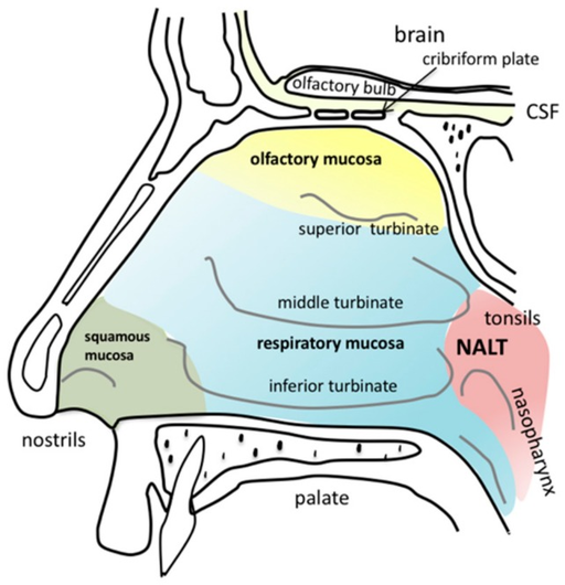
What is the importance of knowing nonmalignant lesions of the inferior turbinate? These cause nasal obstruction and thus need more investigation. Here is the abstract of a review we published.

Abstract
Purpose of review
The inferior turbinates are routinely examined by otolaryngologists on anterior rhinoscopy and nasal endoscopy. Most lesions of the inferior turbinate are benign but can often be confused with malignancy. This review highlights the broad differential of nonmalignant lesions of the inferior turbinates and their management.
Recent findings
A variety of infectious, inflammatory, neoplastic, and vascular lesions may affect the inferior turbinates. The most common nonmalignant lesions of the sinonasal region are nasal polyps, inverted papillomas, hemangiomas, and angiofibromas. Early lesions are often asymptomatic and discovered incidentally on routine examination. As these lesions grow they present with nonspecific signs that can be seen in benign, malignant, and infectious etiologies. The most common signs and symptoms are nasal obstruction, rhinorrhea, epistaxis, sinusitis, and hyposmia. Most nonmalignant lesions have characteristic appearances but definitive diagnosis is achieved with biopsy or culture. If the lesions are small the biopsy itself is often curative.
Summary
Lesions of the inferior turbinates are rarely isolated to these structures alone. Careful examination can non-invasively assist in the early diagnosis of extensive lesions. Once malignancy and processes such as invasive fungal sinusitis or inverted papillomas have been ruled out, treatment of these lesions is ordinarily non-complicated and definitive.
Introduction
Nonmalignant lesions of the sinonasal region are regularly seen by nearly all otolaryngologists. The most common underlying etiologies are nasal polyps (67.6%) followed by inverted papillomas (7.1%), hemangiomas (6.7%), and juvenile nasopharyngeal angiofibromas (JNAs) (5.5%) [1]. A majority of patients affected are males (60.7%) between the ages of 10 and 40 (73.4%) [2]. The inferior turbinates are important structures to consider when evaluating these patients as the structures are routinely and readily visualized on physical examination with anterior rhinoscopy or nasal endoscopy in the office or at the bedside. Moreover, lesions affecting the inferior turbinates are rarely isolated to only these structures and may represent a more extensive process.
Clinically, the most common presenting complaints of nonmalignant lesions of the sinonasal region are nasal obstruction (97.3%), rhinorrhea (49.1%), and decreased sense of smell (31.25%)
[2] Malignant sinonasal masses such as squamous cell carcinoma, adenocarcinoma, and lymphoma may present similarly; however, select signs and symptoms do vary between benign and malignant lesions. For instance, a recent study finds that at the time of presentation sinusitis was associated with benign lesions in 44.4% of cases as opposed to only 7.0% of malignant cases [1]. Also of note, epistaxis or bloody discharge was observed in only 5% of benign cases compared to being present in 76.6% of malignant cases [1]. Other concerning findings that may suggest malignancy are diplopia,
Key Points
-
Lesions of the inferior turbinates are rarely isolated to these structures alone and may be indicative of more extensive processes.
-
The most common nonmalignant lesions of the sinonasal region are nasal polyps, inverted papillomas, hemangiomas, and angiofibromas.
-
Nasal obstruction, rhinorrhea, and sinusitis without epistaxis or bloody nasal discharge are suggestive of nonmalignant lesions affecting the sinonasal region. facial paresthesias, anosmia, proptosis, and cervical lymphadenopathy.
In addition to history and physical examination, imaging and biopsy are critical components in diagnosing nonmalignant lesions of the region. Computed tomography (CT) is extremely useful in evaluating these lesions based on location, enhancement patterns, and bony changes. Although CT findings vary by etiology, a recent study found that benign lesions such as JNAs were often associated with bony deformity or erosion and malignant lesions were more likely to involve bony destruction and complete sinus opacification [3]. MRI can also be useful in select cases due to the advantage of greater soft tissue discrimination. Lastly, definitive diagnosis is often dependent on tissue biopsy. The inferior turbinates’ anterior and easily accessible position is clinically useful for obtaining such biopsies in the outpatient setting with the use of a local anesthetic. Often the lesion can be completely removed with an excisional biopsy.
The differential diagnosis for nonmalignant lesions affecting the inferior turbinate and sinonasal cavity is very broad. In this update, we have organized these processes into four categories: infectious, inflammatory, neoplastic, and vascular. We will focus on manifestations within each grouping, diagnostic findings, and treatment methods. Within the past year, a majority of the literature highlighting lesions specific to the inferior turbinate have been case reports. Many of these reports illustrate rare presentations of already uncommon lesions.
Infectious Lesions
A variety of infectious lesions may affect the inferior turbinates. Potential bacterial causes include Klebsiella rhinoscleromatis leading to rhinoscleroma, Mycobacterium leprae, Actinomycosis, coagulase negative staphylococci, Staphylococcus aureus, and Enterobacter cloacae[4]. Fungal sources to consider are blastomycosis, coccidiomycosis, rhinosporidiosis, mucormycosis, aspergillosis, and Conidiobolus[5]. Polymicrobial infections should also be investigated. For instance, a recent case report by Mahomed et al. [8] highlighted a patient suffering from mucormycosis and Aspergillus coinfections.
In nearly all cases definitive diagnosis is accomplished with culture and biopsy, which is easily obtained in the office due to the accessibility of the inferior turbinate. Systemic empiric antibiotic or antifungal treatment followed by a narrowed antimicrobial is standard for these infectious lesions. Surgical debridement may also be necessary, especially when invasive fungal sinusitis is suspected. Such patients are more likely to be diabetic or immunocompromised and present with facial pain, fevers, black discharge, and necrotic or dusky appearing inferior turbinates [8,9]. Because mucormycosis and the associated invasive sinusitis often begin at the inferior and middle turbinates, early detection and aggressive treatment by otolaryngologists can prevent morbidities associated with advanced disease [1].
Inflammatory Lesions
Many systemic conditions may manifest as lesions of the inferior turbinates. Such processes include: granulomatosis with polyangiitis (GPA), eosinophilic granulomatosis with polyangiitis (EGPA), and sarcoidosis. These diseases may lead to granulation and eventual necrosis in the region [11]. Specifically, a recent study analyzing nasal brush-ings of the inferior turbinates of GPA patients ident-ified 339 genes uniquely expressed in those affected [1,2]. Recent literature also indicates that sarcoid nasal involvement is present in up to 4% of all sarcoidosis cases [11,13]. These findings suggest that the inferior turbinate is often representative of more systemic involvement.
Many other inflammatory etiologies including inhaled substances and foreign bodies causing local inflammation may also damage the inferior turbinates. For instance, cocaine commonly leads to inflammatory complications in addition to the direct effect of the associated micro-trauma when nasally snorted [11]. Oycontin abuse will often cause necrosis and a white appearance to the inferior turbinates, which can be confused with fungus [14]. Additionally non-addressed nasal foreign bodies may lead to rhinoliths leading to local structure compromise [15]. Further, rosacea also leads to local destruction secondary to the effects of inflammatory processes [11].
In many cases, the preferred treatment of non-infectious inflammatory causes of inferior turbinate lesions is based upon steroid and antimetabolite agents. Specifically, glucocorticoids followed by cytotoxic agents in refractory cases are often utilized in treating systemic sarcoidosis, GPA, and EGPA [13]. Additionally, in many cases, such as sarcoidosis limited to the sinonasal region, endoscopic sinus surgery and excision are necessary adjuvant treatments [16].
Neoplastic Lesions
Approximately 70% of all masses arising in the sinonasal region are benign neoplastic lesions whereas malignant and non-neoplastic masses comprise about 20 and 10% of lesions, respectively [1]. Of these benign lesions, inflammatory polyps are by far the most common accounting for nearly 70% of findings in a 10-year retrospective study involving over 600 cases [1]. Inverting papillomas were the second most common benign tumor at approximately 7% followed distantly by a diverse and varied sampling of rare benign lesions [1]. The recent literature describes many of these uncommon lesions, which involve the inferior turbinate and include osteomas, aneurysmal bone cysts, ectopic teeth, schwannomas, and fibrous tumors [17,18,20,21]. Other rare benign masses of note include squamous papillomas, tumors of the minor salivary glands, paragangliomas, and gliomas [22].
CT is recommended in the work-up of such lesions due to their accessibility, cost, and acuity for bony changes. A recent study evaluating the CT findings of histologically confirmed cases of sinonasal neoplasia reveals distinct patterns of benign lesions in the region [3]. For instance inverting papillomas were found to lack calcification in all cases and often contained a focal area of sclerosis [3]. CT is also useful in evaluating or staging malignancy and in future surgical planning if resection is deemed necessary. MRI may also prove useful in evaluating soft tissue and perineural involvement. Distinct patterns on MRI, such as the cerebriform appearance of inverting papillomas, are also useful in evaluating patients. Ultimately MRI is less often utilized due to its associated time and cost.
Biopsy is also necessary in diagnosis and to rule out malignancy. Of note, it is important to identify inverting papillomas due to their propensity to aggressively invade the orbit or cranium and carry a 5–15% risk of malignant transformation [23]. A recent study also found that histologic grading of the inferior turbinate is an effective prognostic indicator of inverting papillomas. Zhao et al. [23] reported on inverting papilloma biopsies taken from the inferior turbinate and concluded that a lack of respiratory epithelium, the prominence of stratified squamous epithelium, and cytoplasmic glycogenation were all histologic features associated with a greater rate of inverting papillomas recurrence. Masses that fit this description, known as grade 2, were found to recur in 95% of cases [23].
The definitive treatment of benign lesions of the inferior turbinate is most commonly with local surgical resection or cautery [17,18,20,21]. Inverting papillomas warrant more aggressive excision and are often removed endoscopically. Medial maxillectomy may be considered in advanced stage inverting papillomas or when masses are less accessible such as when extending to the anterior and inferior walls of the maxillary sinus [23]. Of note, inverting papillomas should be distinguished from squamous papillomas, which visibly present similarly, however, do not carry a risk of malignant transformation [22].
Vascular Lesions
A variety of vascular lesions may also affect the inferior turbinate. The most common etiologies include angiofibromas, hemangiomas, pyogenic granulomas, and angioleiomyomas [24,25,26]. Most angiofibromas are JNAs found in young males; however, recent literature has also described extra-nasopharyngeal angiofibromas (ENPA) involving the inferior turbinates of both genders equally between ages 8 and 67 years old [27,28,29].
Because of the underlying pathogenesis, vascular lesions often present as smooth pink or red masses with recurrent epistaxis. These lesions will often bleed even on routine physical examination. If a mass begins to bleed in the office with an injection of the local anesthetic in preparation for a biopsy, we recommend pursuing the biopsy in the operating room where potential extensive bleeding can be better controlled.
Minor vascular lesions can be managed in the office with local chemical cautery. For more extensive lesions, treatment is with endoscopic surgical excision at the base, cautery, cryotherapy, and laser therapy [1,25,26,30].
Conclusion
Very few lesions of the sinonasal region will be completely isolated to only the inferior turbinates. Because of how routinely examined and accessible these structures are, otolaryngologists can diagnose benign lesions of the inferior turbinate early. Although benign and malignant lesions may both present similarly there are many clear clinical and radiographic indicators to differentiate the two before a biopsy is even taken. Once diagnosed the nose and paranasal sinuses, treatment of these nonmalignant lesions is ordinarily straightforward and definitive with medical or surgical intervention.

